
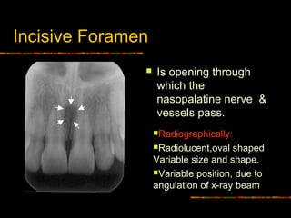
Within this canal lies the nasopalatine nerve and the vascular anastomosis between the greater palatine and sphenopalatine arteries. On periapical x-ray images the incisive foramen is located in the midline between the roots of the central incisors.

Route Of The Incisive Canal Of The Mandible Mic Download Scientific Diagram
Only in a very few radiographs will the incisive canal or nasopalatine canal be.

. Open Journal of Radiology Vol. Coronoid process is the thin triangular-shaped process of the anterosuperior aspect of the ramus. Panoramic radiographs can be used for visualization of the mental foramen and a potential anterior looping but not for locating the mandibular incisive canal.
Panoramic radiographs can be used for visualization of the mental foramen and a potential anterior looping but not for locating the mandibular incisive canal. Accessory Mental Foramen Misdiagnosed as Radiolucent Tumour by Conventional Dental Radiography. International Journal of Dentistry.
Raitz Ricardo Shimura Elisabeth Chilvarquer Israel Fenyo-Pereira Marlene. 30 November 2010 1346 UTC Source Original text. 150 cases with bilateral MIC were analyzed.
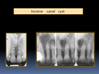
NASOPALATINE duct cysts are cysts which form in the incisor canal region of the maxilla and originate in the nasopalatine duct or its remnants. The anatomy of this area and especially the knowledge for the existence of the MIC is very important for the dentist and the oral surgeon because common surgical procedures performed in this area such as insertion of. Our goal is to evaluate identification of MIC by both panoramic radiograph PAN and cone-beam computed tomography CBCT.
An anatomical variation to be considered is the anterior looping of the mental nerve in 11 of images. Assessment of the Mandibular Incisive Canal by Panoramic Radiograph and Cone-Beam Computed Tomography. The mandibular incisive canal mental foramen and associated neurovascular bundles exist in different locations and possess many variations.
2014 Issue 2014 31 Dec. The incisive canal located at the midline posterior to the central incisor is an important anatomic structure of this area to be considered while planning for immediate implant placement in maxillary central incisor region. However complications may arise due to an extension anterior to the mental foramen that forms the mandible incisive canal MIC.
2014 pp1-6 6 p. The incisive canal also known as the nasopalatine canal is an interosseous conduit through the anterior maxilla connecting the oral and nasal cavities. The pear-shaped radiolucency between the apices of the central incisors can be mistaken for periapical pathology or cyst formation.
The purpose of the present study is to assess incisive canal characteristics using CBCT sections. Mean canal length was 1863 235 mm and males have significantly longer incisive canal than females. In addition the angulation of the X-ray beam in panoramic radiography is about 78 from below.
Its appearance is quite variable due to normal anatomic variation and due to the operators angulation of the x-ray beam. An incisive canal was identified in 15 of the images with good visibility in only 1. Our goal is to evaluate identification of MIC by both panoramic radiograph PAN and cone-beam computed tomography CBCT.
The region between mental foramens is considered as a zone of choice for implants. The incisive foramen generally appears in most panoramic radiographs though not with the clarity seen in periapical radiographs. Results The incisive canal was found in 87 of the scans.
This results in some distortion of the actual mandibular anatomy and may lead to misinterpretation. However complications may arise due to an extension anterior to the mental foramen that forms the mandible incisive canal MIC. Assessment of the Mandibular Incisive Canal by Panoramic Radiograph and Cone-Beam Computed Tomography.
It can be single or multiple. International Journal of Dentistry Vol. These cysts have no direct relationship to the teeth but in their growth may encroach upon the incisor apices.
It is located in the maxilla in the incisive fossa midline in the palate posterior to the central incisors at the junction of the medial palatine and incisive sutures. It suggests that the clinicians should carefully identify these. Research Article Assessment of the Mandibular Incisive Canal by Panoramic Radiograph and Cone-Beam Computed Tomography Ricardo Raitz12 Elisabeth Shimura3 Israel Chilvarquer34 and Marlene Fenyo-Pereira4 1 Discipline of General Pathology University of Sao Caetano do Sul USCS Sao Caetano do Sul SP.
However complications may arise due to an extension anterior to the mental foramen that forms the mandible incisive canal MIC. Contribs created this work entirely by myself color006400Contribs. The incisive foramen also known as nasopalatine foramen or anterior palatine foramen is the oral opening of the nasopalatine canal.
This might be explained by the fact that the incisive canal is less corticalized and has a smaller diameter than the mandibular canal. Periapical radiograph depicting the junction of the mandibular canal and the mandibular incisive canal near the mental foramen. Assessment of the mandibular incisive canal by panoramic radiograph and cone-beam computed tomography Objectives.
The mean width of bone anterior to the incisive canal was 632 143 mm. As age of the subjects increased incisive foramen diameter and incisive canal length were found to be increased. Individual gender age race assessing technique used and degree of edentulous alveolar bone atrophy largely influence these variations.
The mandibular incisive canal MIC is described as an anterior extension of the mandibular canal anterior to the mental foramen containing a neurovascular bundle. The region between mental foramens is considered as a zone of choice for implants. The mean endpoint was approximately 1098 and 1026 mm anterior to the mental foramen for left and right side respectively without a.
To verify its existence for.

A Panoramic Radiograph Shows The Anterior Loop And Incisive Canal Download Scientific Diagram

The Radiology Of Developmental Dental Defects Demystified An E Based Learning System Intechopen

Pdf The Evaluation Of Visibility Of Mandibular Anatomic Landmarks Using Panoramic Radiography Semantic Scholar

6 Essentials Of Dental Radiographic Analysis And Interpretation Pocket Dentistry

Normal Radiographic Anatomical Landmarks

Mouth Incisive Canal Cyst Professional Radiology Outcomes

6 Essentials Of Dental Radiographic Analysis And Interpretation Pocket Dentistry

Opg Showing Incisive Foramen And Mental Foramen Download Scientific Diagram
Intra Oral Radiographic Anatomical Landmarks
Normal Radiographic Anatomical Landmarks
Periapical Radiograph 1 Year After Treatment Bone And Teeth Showing Download Scientific Diagram

Maxillary Anterior Landmarks Intraoral Radiographic Anatomy Continuing Education Course Dentalcare Com

Cone Beam Ct Images Showing A B Sagittal Cuts Of A Bifid Incisive Download Scientific Diagram

Figure 2 From Visibility Of Mandibular Anatomical Landmarks In Panoramic Radiography A Retrospective Study Semantic Scholar

Xmlinkhub

Figure 1 From Visibility Of Maxillary And Mandibular Anatomical Landmarks In Digital Panoramic Radiographs A Retrospective Study Semantic Scholar

Radiographic X Ray Taken After Root Canal Treatment On Tooth 12 A Download Scientific Diagram

Maxillary Anterior Landmarks Intraoral Radiographic Anatomy Continuing Education Course Dentalcare Com

Anterior Loop Of The Mental Nerve And Its Radiologic Imaging A Review Semantic Scholar